Hoof Anatomy

You are currently browsing articles tagged Hoof Anatomy.

Mustangs from American WestInterested in feral horse hoofs as a model for the domestic horse?

Check out this post from Fran Jurga’s Hoof Blog, featuring links to a video and 46-page research report from researchers Chris Pollitt and Brian Hampson – detailing their work with and findings about the hoofs of brumbies – the feral horses of Australia.

Tags: , ,

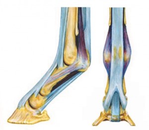
Diagram of Equine Lower Leg, showing Suspensory Ligaments

Suspensory ligament (above fetlock) shown in dark purple. Anterior branches of suspensory ligament (below fetlock) are light purple.

Read up on equine suspensory ligament injuries and anatomy in the free downloadable 20-page booklet ‘Suspensory Ligament Injuries in Horses’ from the UC Davis School of Veterinary Medicine – written for horse owners and for horse professionals.

The booklet is generously illustrated with drawings, photos, and ultrasound images showing the anatomy and function of the equine suspensory apparatus in health and in injury. Read the rest of this entry »

Tags: , , , ,

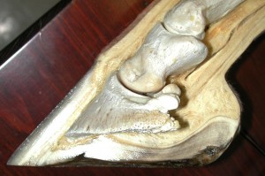
Horse Hoof Anatomy - Cross section view

Check out this amazing database of equine anatomy photos from the Cornell University College of Veterinary Medicine, designed to accompany ‘Rooney’s Guide to the Dissection of the Horse’.

Please note: The images in the database are photographs of dissected cadavers. The database and images associated with these pages are intended for use by veterinary professionals and may not be suitable for all individuals; parental guidance is advised.

Enter the Cornell University College of Veterinary Medicine Equine Anatomy Photo Database

To begin browsing the database, click the large red button labeled ‘Search the Database’ on the home page of the database.

For anatomy photos of the equine hoof and lower leg – choose [Chapter 6: Forelimb] from the Select Chapter drop-down menu.

Tags:

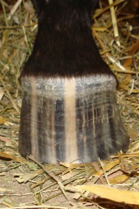
front view of a horse's hoof

Is this a normal, healthy hoof? This hoof is not symmetrical - the hoof wall on the left side of the photo is more upright, while the wall on the right side of the photo is slanted outward.

Author, veterinary pathologist, and  authority on equine biomechanics and anatomy Dr. James Rooney, DVM, says yes – and no.

Left and right hooves are often symmetrical images of each other mirrored across the center line of the horse’s body, yet are not symmetrical across their own center lines.

Look at the photo of the hoof on the left. Is this a right hoof, or a left hoof? (Answer below.) The inside wall of most horse hooves appears steeper, while the outside wall has a more gradual slope.

Read the rest of this entry »

Tags: , , , ,

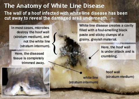
The photo below shows a closeup view of a hoof with a fairly advanced case of white line disease. The infection has destroyed the inner unpigmented layer of the hoof wall, leaving behind a v-shaped slot between the dark-colored outer wall and the yellowish white line.

Because it is difficult to medicate the active disease at the top of such a deep cavity, the hoof wall has been cut away and diseased tissue has been removed.

labeled photo of hoof infected with white line disease

White line disease is an infection that can attack and destroy large areas of your horse’s hoof wall.

Click to view larger photo.

Tags: , ,

« Older entries